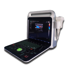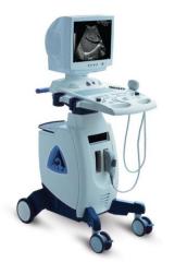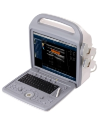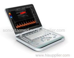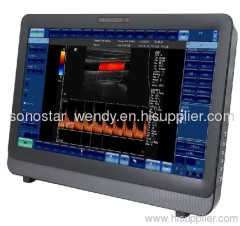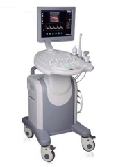
Color Doppler Ultrasound System
| Min. Order: | 1 Piece/Pieces |
|---|---|
| Payment Terms: | L/C, D/P, D/A, T/T, WU |
| Supply Ability: | 10000pcs/month |
| Place of Origin: | Zhejiang |
Company Profile
| Location: | Ningbo, Zhejiang, China (Mainland) |
|---|---|
| Business Type: | Manufacturer, Trading Company |
Product Detail
| Means of Transport: | by seasby air or by express courier |
|---|---|
| Brand Name: | OEM |
| Production Capacity: | 10000pcs/month |
| Packing: | carton |
| Delivery Date: | 7 days |
Product Description
color Doppler ultrasonic diagnostic system is composed of advanced technology of digital color Doppler and wide frequency probe, internet applications system with general measuring function, kinds of leading image process technology, which reaches to leading level in the world. It is whole-body applied digital color ultrasonic diagnosis system of Haiying ultrasonic family with clear image.
Features
Applications
● Abdominal ● Small Parts ● Obstetrics
● Gynecological ●Cerebrovascular
● Peripheral Vascular ● Cardiac
Applications
● Abdominal ● Small Parts ● Obstetrics
● Gynecological ●Cerebrovascular
● Peripheral Vascular ● Cardiac
Imaging Modes
●3D ● Color Doppler
● Energy Doppler ● Spectrum Doppler
● M Mode ● Dual Image Mode
● Duplex, 3D & PW Doppler
● Triplex, 3D, Color & PW Doppler
●3D ● Color Doppler
● Energy Doppler ● Spectrum Doppler
● M Mode ● Dual Image Mode
● Duplex, 3D & PW Doppler
● Triplex, 3D, Color & PW Doppler
System Description
● The system is a 128 channel, high resolution ultrasound imaging system with patented Adaptive Focusing which enables high performance ultrasound imaging from 2MHz to 10MHz.
● Each transducer has three imaging frequencies and two or three Doppler frequencies available.
● The system has sophisticated report generating capabilities for OB, Vascular, Cardiac, Gynecology and Urology.
● This PC based system can store images and reports to disk. Images and report can be copied to external media. HTML versions of reports can easily be copied, emailed and viewed by any Internet Browser.
● The system provides multiple application specific presets for each probe. Custom presets can be generated by the user.
● Windows 2000 Professional Operating System enables support of a wide range of peripherals and is readily upgraded with software enhancements.
Hardware and Software
● 128 Element Linear and Curved Arrays
● 2 Transducers can be connected electronically selectable
● Receiver Dynamic Range > 125dB
● Total Dynamic Range > 150dB
● Display Dynamic Range > 70dB
● Eight Transmit Focal Zones for high resolution throughout the image
● Continuous dynamic focusing on receive
● Center frequency range from 2.5MHz to 9MHz (band edge from 1.5MHz to 12MHz)
● Multi-frequency operation for all probes
● B Mode Imaging frame rates up to 34 frames per second
● B Mode line density up to 512 lines
● Field of View (FOV), variable, up to 25 cm in five steps
● Variable transmit power and variable image gain
● Slide pot TGC controls
Control Panel and User Interface
● Ergonomic control panel with controls organized by mode
● Alphanumeric QWERTY keyboard
● Trackball with Set and Esc keys
● Integrated stereo speakers
● 3D image controls: Power, Gain, TGC, Depth, Focus, Magnify, Zoom, Dual, Orientation
● Image Enhancement: Dynamic Range, Persistence, Gray Scale maps, Edge Enhancement
● Doppler controls: Angle/Steer (linear arrays), PRF (velocity range), Angle Correction, Baseline Shift, Gain ,Power
● Color controls: Velocity mode, Power Mode, Color priority, Color frame rate, Color maps, Color persistence
● Patient data entry
● Image Acquisition: Cine review, Image storage, Cine storage
● Image and Report retrieval
● Image Annotation
Monitor
● 15 inch, color VGA
● Swivels, Tilts
● Brightness, Contrast, and Color temperature controls
● 15 inch, color VGA
● Swivels, Tilts
● Brightness, Contrast, and Color temperature controls
Imaging Display
● Dual image display
● Image orientation control, both horizontal and vertical
● Magnify up to 3×
● 2× Zoom, with full screen image and magnified section
● Variable Sector Angle for curved array transducers
● 256 Gray shades
● Eight gray scale maps
● Display dynamic range from 30 – 70 dB
● Display of output power
● Display of TI and MI (Track Ⅲ) in all modes
● Variable persistence
● Edge enhancement
● Image storage for more than 10000 frames on local drive (unlimited with external media)
● Cine Loop:
Stores up to 256 frames of B & W or Color images (with optional 512MB memory, 128 frames standard)
Trackball control of frame-by-frame image selection
Controls for cine play back
Controls for trimming
Cine loops can be saved and retrieved as part of patient record
● Measurement capability in B Mode:
Distance
Area by ellipse
Area by trace
Curve length
Volume (required two images)
Angle
● Full image annotation capabilities including:
Text entry and editing
Pre-programmed vocabulary (user definable)
Arrows and pointers
Body marks that cover many applications and orientations
M Mode
● B Mode & M Mode display
● Full screen M Mode display
● Variable scroll speed 2, 3, 4, 5, 6, 8 seconds
● User adjustable M Mode line
● Time, velocity, acceleration, Heart Rate measurements
● Variable Dynamic Range and Persistence
● B Mode & M Mode display
● Full screen M Mode display
● Variable scroll speed 2, 3, 4, 5, 6, 8 seconds
● User adjustable M Mode line
● Time, velocity, acceleration, Heart Rate measurements
● Variable Dynamic Range and Persistence
PW Doppler
● Duplex (simultaneous) 2D and Doppler
● Triplex mode, 2D, Color & PW
● Steered mode for linear arrays
● Wide range of velocities (PRFs)
● Variable wall filters
● Adjustable gate size from 1 to 7 mm
● Doppler angle correction for velocity measurements
● Baseline shift and invert functions
● Variable spectral sweep speeds
● Automatic mean and peak velocity traces
● Variable gray maps
● Measurement capability in PW Mode: Time, Velocity, Acceleration
Color Doppler Imaging
● Region of Interest (ROI), size and position variable
● Steered Color with linear arrays
● Variable wall filters
● Variable Color priority
● Multiple Color maps
● Variable Color frame rate
● Velocity and power displays
● Variable color persistence
● Color invert
● Region of Interest (ROI), size and position variable
● Steered Color with linear arrays
● Variable wall filters
● Variable Color priority
● Multiple Color maps
● Variable Color frame rate
● Velocity and power displays
● Variable color persistence
● Color invert
Power Doppler Imaging
● Region of Interest (ROI), size and position variable
● Steered color with linear arrays
● Variable wall filters
● Variable Color priority
● Multiple Color maps
● Variable Color frame rate
● Variable color persistence
● Region of Interest (ROI), size and position variable
● Steered color with linear arrays
● Variable wall filters
● Variable Color priority
● Multiple Color maps
● Variable Color frame rate
● Variable color persistence
Calculation Packages
All measurement packages support a powerful Worksheet capability that enables review and manipulation of measurement information.
All measurement packages support a powerful Worksheet capability that enables review and manipulation of measurement information.
Obstetrics Measurements
● Calculation of Estimated Gestational Age (EGA) based upon: AC, BPD, BD, CRL, FL, GSD, HC, HL, MAD, OFD, TL, UL
● Calculation of Estimated Fetal Weight (EFW)
● Calculation of growth parameter ratios
● Graphic display of statistical growth charts
● Biophysical Score report
● Structural Report
● Calculation of Estimated Gestational Age (EGA) based upon: AC, BPD, BD, CRL, FL, GSD, HC, HL, MAD, OFD, TL, UL
● Calculation of Estimated Fetal Weight (EFW)
● Calculation of growth parameter ratios
● Graphic display of statistical growth charts
● Biophysical Score report
● Structural Report
Cardiac Measurements
● Cardiac index
● Cardiac output
● LV end-diastolic volume
● LV end-systolic volume
● Ejection fraction
● Stroke index
● Stroke volume
● IVS/LVPW ratio
● Aortic valve pressure gradient
● Aortic pressure half time
● Aortic valve area
● Mitral valve area
● Left ventricular outflow tract pressure gradient
Vascular Measurements
● Time Average Mean Velocity (TAM)
● Time Average Max Velocity (TAMX)
● Pulsatility Index (PI)
● Resistive Index (RI)
● Peak Systolic Velocity (PSV)
● End Diastolic Velocity (EDV)
● S/D Ratio (PSV/EDV)
● PSV_ICA/PSV_CCA
● Velocity Ratio (Velocity 1 / Velocity 2)
● % Stenosis by Diameter
● % Stenosis by Area
Focusing
Transmitting focusing (8 ranges), receiving continuous fousing
Transmitting focusing (8 ranges), receiving continuous fousing
Display Prameter
Acoustic power, total gain, dynamic range, TGC curve, gain of Doppler, pulse repetition frequency, wall-filtering ratio.
Acoustic power, total gain, dynamic range, TGC curve, gain of Doppler, pulse repetition frequency, wall-filtering ratio.
Image Process
Colorful map, frame average processing, angle adjustment and wall filter
Colorful map, frame average processing, angle adjustment and wall filter
Gray 256
Image Storage
Storage and playback of 128 continuous image
Image Output
Output to floppy disk, USB and color video printer
Storage and playback of 128 continuous image
Image Output
Output to floppy disk, USB and color video printer
Standard Configuration
Main unit,15"RGB color monitor,3.5MHz 60R65D multi-frequency convex probe
7.8MHz38L multi-frequency linear probe,6 USB ports, DVD-RW, 2 probe connectors
Main unit,15"RGB color monitor,3.5MHz 60R65D multi-frequency convex probe
7.8MHz38L multi-frequency linear probe,6 USB ports, DVD-RW, 2 probe connectors
Option
3.3MHz72D20R multi-frequency convex probe
7.0MHz14D10R multi-frequency convex probe
Color video printer
3.3MHz72D20R multi-frequency convex probe
7.0MHz14D10R multi-frequency convex probe
Color video printer