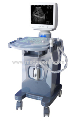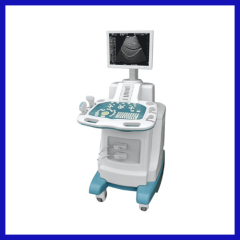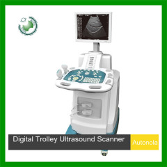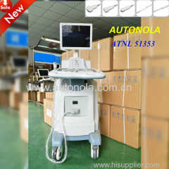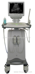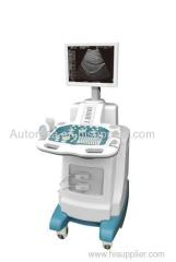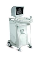
Trolley Ultrasound Scanner
| Place of Origin: | Zhejiang |
|---|
Company Profile
| Location: | Ningbo, Zhejiang, China (Mainland) |
|---|---|
| Business Type: | Manufacturer, Trading Company |
Product Detail
| Model No.: | TH-3000G2 |
|---|---|
| Brand Name: | OEM |
Product Description
Full Digital Ultrasonic Diagnostic Equipment
Leading Digital Technology
DBF:Digital Beam Former
RDF:Real-time Dynamic Filtering
DFS:Dynamic Frequency Scanning
RDA:Real-time Dynamic Aperture
DBF:Digital Beam Former
RDF:Real-time Dynamic Filtering
DFS:Dynamic Frequency Scanning
RDA:Real-time Dynamic Aperture
Features:
Trolley design
14 inch high-resolution monitor
8 TGC controls
High-quality images
Broadband multi-frequency probes
Cineloop
Permanent image storage
USB port
14 inch high-resolution monitor
8 TGC controls
High-quality images
Broadband multi-frequency probes
Cineloop
Permanent image storage
USB port
Imaging Processing:
- Planar images
- The functions of frame correlation, γ correction and edge enhancement held, as well as the functions of pretreatment, post processing, left-eight reversing and up-down reversing
- Image displaying ratio: step-less magnification which can be increased. Real-time local zooming held
- Maximum storage of more than 100,000 images. Images recorders optional
- Case history created and created automatically
- Images manage system, Case history database system and diagnosis & measurement pre-treat system
Technical Specification:
Display | 14 inch high-resolution monitor |
Scanning mode | Electronic linear array, electronic convex linear |
Display mode | B, B/B, B/M, M, 4B |
Magnification | ×1.0, ×1.2, ×1.5, ×2.0 |
Gray Scale | 256 level |
Displaying depth | 240mm, and depth could be increased and stepless magnified |
Resolution | Horizontal: ≤2mm Vertical: ≤1mm |
Geometrical precision | ≤5% |
Blind area | ≤3mm |
TGC | 8-section TGC adjust |
Image Polarity | left/right turn, positive /negative turn and up / down turn |
Cineloop | successive 256 frames |
Output Interface | two output interfaces, VGA and PAL video signal output |
Power Supply Range | AC 110V 60Hz AC 220V 50Hz |
Focusing modes | dynamic-emitting focus, dynamic-receiving focus, dynamic aperture, adaptive acoustic lens |
Output interface | AV, S-video, VGA |
USB connector | Yes |
Measurement and Analysis Software:
- The General Measurement and Analysis
- The Measurement and Analysis for Gynecology
- The Measurement and Analysis for Obstetrics
- The Measurement and Analysis for Urology
- The Measurement and Analysis for Small parts
- The Measurement and Analysis for Heart and the ambient vessels
Remarks: for each picture there are more than 5 parameters for reference, such as distance, area, perimeter, angle, volume, gestational age, fetus' weight, heart rate, slope rate, flux, left ventricle and so on
Standard Configuration:
- Main unit
- 14 inch monitor
- 256-frame cineloop
- Images permanent image storage
- Two probe connectors
- USB port
- 3.5MHz electronic convex transducer (2.5/5.0MHz)
Optional:
- Superconductivity Visual Probe
- Electronic transvaginal transducer (5.0/7.5MHz)
- Electronic High-frequency linear transducer (6.5/8.5MHz)
- MSF: Mass storage
- Jet printer, laser printer, video printer
- DICOM 3.0 interface


