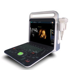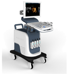
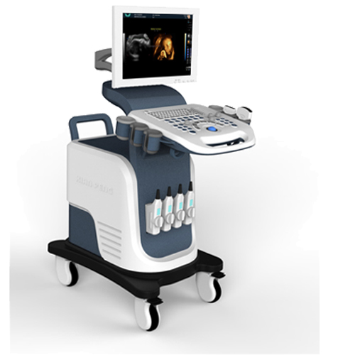
Real-time 4D imaging color ultrasonic diagnosis system
| Min. Order: | 1 Set/Sets |
|---|---|
| Trade Term: | FOB,CFR,CIF,CIP,CPT,FCA |
| Payment Terms: | Paypal, L/C, T/T, WU, Money Gram |
| Supply Ability: | 2000 |
| Place of Origin: | Sichuan |
Company Profile
| Location: | Mianyang, Sichuan, China (Mainland) |
|---|---|
| Business Type: | Manufacturer, Trading Company, Service |
Product Detail
| Model No.: | XF7800(4D) |
|---|---|
| Means of Transport: | Ocean, Air, Land |
| Brand Name: | XianFeng |
| Ultrasound scanner: | Mobile ultrasound |
| USG: | Doppler scan |
| 3D fetus imaging: | Ultrasound machine |
| Power doppler ultraound: | Cardiac ultrasound |
| Multipurpose doppler: | 4D abdominal ultrasound |
| Vascular ultrasound: | Ultrasonography |
| Abdomen Ultrasound: | Trolley Doppler |
| 3D doppler scan: | Clinical ultrasound |
| Production Capacity: | 2000 |
| Packing: | 1090mm*700mm*1130mm |
| Delivery Date: | within 7 days |
Product Description
XF7800(4D) full digital color doppler ultrasound diagnostic system is based on Windows,stable and easier operation.high accuracy and clear imaging.It support 4D and triple real-time imaging.The 4D imaging of this model adopts the current advanced synchronous driven volume probe and 4D software, and stabilizes the image scanned by the probe in real time. It can observe the facial expressions of the fetus in real time and leave precious growth record video for people.It is widely used in abdominal,gynecology,obstetric,pediatric,urology,andrology,small organ and musculoskeletal etc.
XF7800(4D)Real-time 4D imaging color ultrasonic diagnosis system

* B,B+B,4B mode
* B+M mode
* CFM color Doppler mode
* B+CFM mode
* PDI Power Doppler Mode
* B+PDI mode
* PW mode
* Trapezoidal imaging
* Imaging mode(2D,Optional 3D, Optional 4D)
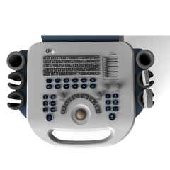
--Application mode: abdomen, gynecology, obstetrics, superficial organ, urologist, heart and user defined model 1-4, total ten models;
--Image mode: digital beam forming, tissue harmonic imaging;
--Acoustic output: Mechanical index and thermal index real-time display;
--Acoustic power:Step is adjustable, real-time display;
--Gray scale: 256 scales;
--Depth display: ≥250mm;
--B/D dual-purpose: linear array: B/PWD; convex array: B/PWD;
--Pseudo color processing: 16 kinds of pseudo color encoding can optional;
--Gain adjusts: 8 segments TGC, B/M/D/C is independently adjustable; TGC curve can show and hide automatically;
--Image magnification: picture in picture zoom in and zoom part function;
--Image processing: Edge enhancement: Multilevel adjustable
--Frame average: Multilevel adjustable
--Line average: Multilevel adjustable
--Focus Optimization: Multilevel adjustable
--Gray Restrain: Multilevel adjustable
--Gamma correction: Multilevel adjustable
--Contrast: Adjustable
--Brightness: Adjustable
--Self-motion optimize function: Built-in multiple check type, according to different inspection organs, preset best image check condition, reduce the adjusting operation keys;
--One-click optimization function: preset several parameters adjusting focus on a Key, a key to realize image fast optimization;
--Measurement and calculation: B mode routine measurement:Distance, circumference, area, volume, angle, ratio, and stenos rate.
--M mode routine measurement: Heart rate, time, distance, speed, ratio, etc.
--Gynecology measurement: Uterus, cervix, endometrial, ovary, follicular.
--Obstetrics measurement: EGA, ETD, fetal weight estimation, AFI index, OB report (including OB tables).
--Cardiology measurement: LV measurement.
--Urology measurement: Prostate volume, displacement volume, bladder capacity, and residual urine output.
PW measurements: Time, speed, Heart Rate, RI, PI, etc.
--Other measurement: Slice volume measurement, hip joint angle measurement.
--Image storage: Image storage, video storage, cine loop, disk storage capacity≥320G;
--Patient data: Medical record management, report inquiry and printing, image video output( HDD ,USB,Optional DVD-RW),built-in ultrasound workstation;
--Reporting system: automatic report generation system, and can be full screen characters in both Chinese and English editor;
--Output interface: USB,DICOM interface;
XF7800(4D)Real-time 4D imaging color ultrasonic diagnosis system
Main Technical Indexes
The performance requirements of gray-scale imaging mode
The color ultrasonic at the gray-scale imaging performance mode should comply with the provisions of the table 1.1
Table 1.1 At the Gray-scale imaging mode the performance of the probe
performance indexes | probe type and nominal frequency | |||
2.0≤f<4.0 | 2.0≤f<5.0 | 5.0≤f<8.0 | 5.0≤f<12.0 | |
a) probe type and model | phased array (type TP16A) | Convex array (type TC60A) | Cavity (type TC10A) | Linear array (type TL40A) |
b) nominal frequency (MHz) | 3.0 | 3.5 | 6.5 | 7.5 |
c) Scan depth(mm) | ≥140 | ≥160 | ≥40 | ≥50 |
d) Lateral resolution (mm) | ≤3(depth≤80) ≤4(80<depth≤130) | ≤3(depth≤80) ≤4(80<depth≤130) | ≤2(depth≤30) | ≤2(depth≤40) |
e) Axial resolution (mm) | ≤2(depth≤80) | ≤2(depth≤80) ≤3(80<depth≤130) | ≤1(depth≤40) | ≤1(depth≤50) |
f) Blind area (mm) | ≤7 | ≤5 | ≤4 | ≤3 |
g) Transverse geometry precision (%) | ≤20 | ≤15 | ≤10 | ≤10 |
h) Longitudinal geometric location accuracy (%) | ≤10 | ≤10 | ≤5 | ≤5 |
i) Slice thickness (mm) | ≤5 | ≤5 | ≤5 | ≤5 |
j) Perimeter and area measured deviation (%) | ≤±20 | ≤±20 | ≤±20 | ≤±20 |
k) M mode time display error (%) | ≤±10 | ≤±10 | ≤±10 | ≤±10 |
Overall dimensions:
Overall dimensions:785mm×586mm×870mm
Net weight:almost 35㎏