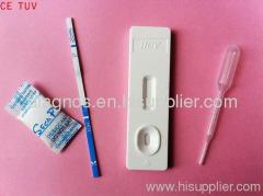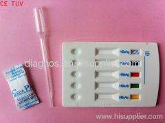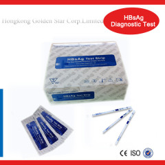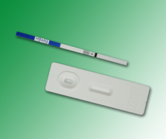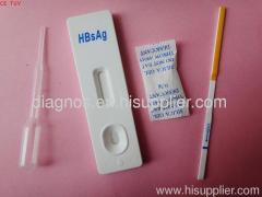
DIAGNOS HBsAg Test/ hepatitis test
| Payment Terms: | T/T, WU |
|---|---|
| Place of Origin: | Jiangsu |
Company Profile
| Location: | Nantong, Jiangsu, China (Mainland) |
|---|---|
| Business Type: | Manufacturer, Trading Company |
| Main Products: | Rapid Test Kits |
Product Detail
| Means of Transport: | Ocean, Air, Land |
|---|---|
| Brand Name: | DIAGNOS/ OEM |
Product Description
A rapid, one step test for the qualitative detection of Hepatitis B Surface Antigen (HBsAg) in serum or plasma.
For professional in vitro diagnostic use only.
INTENDED USE
The HBsAg One Step Hepatitis B Surface Antigen Test Strip (Serum/Plasma) is a rapid chromatographic immunoassay for the qualitative detection of Hepatitis B Surface Antigen in serum or plasma.
SUMMARY
Viral hepatitis is a systemic disease primarily involving the liver. Most cases of acute viral hepatitis are caused by Hepatitis A virus, Hepatitis B virus (HBV) or Hepatitis C virus. The complex antigen found on the surface of HBV is called HBsAg. Previous designations included the Australia or Au antigen. The presence of HBsAg in serum or plasma is an indication of an active Hepatitis B infection, either acute or chronic. In a typical Hepatitis B infection, HBsAg will be detected 2 to 4 weeks before the ALT level becomes abnormal and 3 to 5 weeks before symptoms or jaundice develop. HBsAg has four principal subtypes: adw, ayw, adr and ayr. Because of antigenic heterogeneity of the determinant, there are 10 major serotypes of Hepatitis B virus.
The HBsAg One Step Hepatitis B Surface Antigen Test Strip (Serum/Plasma) is a rapid test to qualitatively detect the presence of HBsAg in serum or plasma specimen. The test utilizes a combination of monoclonal and polyclonal antibodies to selectively detect elevated levels of HBsAg in serum or plasma.
PRINCIPLE
The HBsAg One Step Hepatitis B Surface Antigen Test Strip (Serum/Plasma) is a qualitative, lateral flow immunoassay for the detection of HBsAg in serum or plasma. The membrane is pre-coated with anti-HBsAg antibodies one the test line region of the strip. During testing the serum or plasma specimen reacts with the particle coated with anti-HBsAg antibody. The mixture migrates upward on the membrane chromatographically by capillary action to react with anti-HBsAg antibodies on the membrane and generate a colored line. The presence of this colored line in the test region indicates a positive result, while it's absence indicates a negative result. To serve as a procedural control, a colored line will always appear in the control line region indicating that proper volume of specimen has been added and membrane wicking has occurred.


