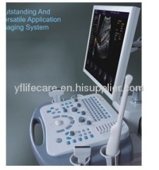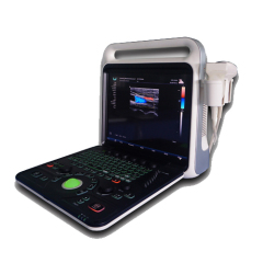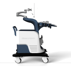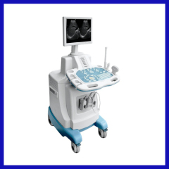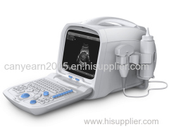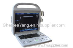
Color Doppler Ultrasonic Diagnostic System
| Min. Order: | 1 UNIT |
|---|---|
| Payment Terms: | Paypal, T/T, WU |
| Supply Ability: | 5/week |
| Place of Origin: | Hebei |
Company Profile
| Location: | Qinhuangdao, Hebei, China (Mainland) |
|---|---|
| Business Type: | Manufacturer, Trading Company, Distributor/Wholesaler, Other |
| Main Products: | Fingertip Oximeter, Pocket Fetal Doppler Color LED Display, Fingertip Pulse Oximeter, Pocket Fetal Doppler, Pulse Oximeters |
Product Detail
| Model No.: | CMS1900 |
|---|---|
| Means of Transport: | Air |
| Display mode: | B, B/B, 4B, B/M, CF, PDI/DPDI, PW, THI |
| Production Capacity: | 5/week |
| Delivery Date: | 2 working days after payment is confirmed |
Product Description
Introductions
CMS1900 is a color doppler ultrasonic diagnostic system with high resolution which possesses powerful computer processing flat. The system is mainly suitable for the diagnosis of abdomen, cardiology, peripheral vessels, gynecology, small organs, urology, muscle,and incretion etc. It adopts doppler ultrasound imaging, digital beam-forming, tissue harmonic imaging (THI), image speckle-reduction and many other advanced image processing technologies, and full digital integrative managing system.The clinic diagnostic requirement can be satisfied by the professional measurement software packages which are set in the system.
Main Features
◆Display mode:B, B/B, 4B, B/M, CF, PDI/DPDI, PW, THI
◆It adopts continuous dynamic receiving aperture (CDA), continuous dynamic receiving focusing (CDF) technologies, which improve the resolution of images.
◆Clear layout for functional keyboard and humane care design make the system easy to operate.
◆It adopts advanced image processing technologies, such as: frame correlation, wall filter, color code figure,image enhance.
◆Scientific probe match: It can avoid wastage of ultrasonic energy, accordingly improve detection capability and image definition.
◆Advanced probe technology:It supports multi-frequency probe, and can adjust to the needed depth of penetration through the probe frequency conversion, so as to satisfy the diagnosis demand of different sufferer.
◆High effective doppler technology: Doppler frame correlation, doppler apace optimize, transmit coding control.
◆Expanded interface:VGA, VIDEO, USB2.0, RJ-45
◆Power supply:AC 100 V~240 V, 50 Hz/60 Hz
Application
Abdomen, obstetrics, gynecology, small organs, urology, incretion, blood vessel/peripheral vessels,cardiology etc.
Main Performance
B Mode
Image invert:up/down, left/right
8 segments TGC control
Dynamic Range, image enhance, frame correlation, gray scale, image rejection can be adjustable.
Cine loop: ≥ 500 frame
Automatically review, Plus/contrary review with single pace, segment review, cine memory.
M Mode
Sampling line speed, dynamic range, gray scale, image rejection can be adjustable.
CF Mode
Pulse repeated frequency adjustable
Color PRI
Color Threshold control
Color baseline control
Doppler frequency selection
Color frame average
Color Transparency
PDI Mode
Linear doppler angle adjustable
Color PRI
The size and position of sampling box adjustable
Image frame correlation, frame frequency adjustable
Wall filter adjustable
PW mode
The size and position of sampling box can be adjusted, doppler frequency conversion
Wall filter adjustable
Three modes (B+CFM/PDI+PWD)
Doppler SV range can be regulated
Support doppler angle correction
Base line move
Color frequency spectrogram sweep speed can be regulated
Can automatically obtain the envelope of frequency spectrum graph
Volume in PW mode can be regulated
Interface language
Support Chinese, English language interface display and input
Image processing
Tissue harmonic imaging, color code imaging, angle steer, frame average processing, image enhance, image rejection, wall filter.
Image storage: Large quantity of memory function.
Image output: To the native disk, mobile hard disk and printer.
Measurement and analysis
Special software as routine measurement and obstetrics, gynecology, abdomen, urology, incretion, blood vessel, cardiology etc.
Generic measurement (beeline distance, curve length, ellipse measuring method or track measuring method to measure area, volume and angle etc.)
Obstetric measure software package
Gynecology measure and calculation software package
Urology measure and calculation software package
Doppler blood flow measurement and analysis
Peripheral vessels measurement and analysis
Standard Configuration
Main Unit
3.5MHz Convex Probe
9.0 MHz Linear Probe
Power Cable
Fuse
Coupling Gel
User Manual
Packing List
Inspection Report
Optional Configuration
3.5 MHz Micro-Convex Probe
6.5 MHz Transvaginal Probe
7.5 MHz Transrectal Probe
7.5 MHz Linear Probe
Monitor
Laser printer, ink jet printer, video printer.
Probe puncture support


