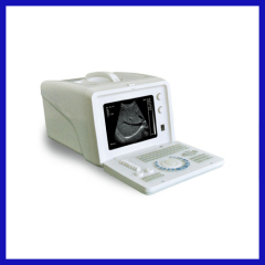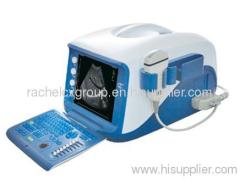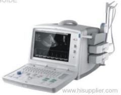
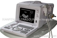
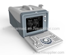
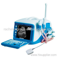
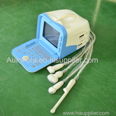
Portable ultrasound scanner
| Supply Ability: | 1000 sets/year |
|---|---|
| Place of Origin: | Hubei |
Company Profile
| Location: | China (Mainland) |
|---|---|
| Business Type: | Manufacturer |
Product Detail
| Model No.: | ZQ-6600plus |
|---|---|
| Brand Name: | OEM or Zoncare |
| Production Capacity: | 1000 sets/year |
| Packing: | carton |
| Delivery Date: | 7 days |
Product Description
Clinical Application and Main Specification
Windows XP Embedded operation system
Three-Dimensional Imaging technology, rebuilt the 3D image from the 2D image.
Application: abdomen, gynecology, obstetrics, urological, small parts and blood vessel.
Automatic measurement software of OB/GYN, urology, pediatrics, cardiology and small parts.
Select different diagnostic measurement formula for different race
Scanning model: Convex, Linear, and Micro-convex
Imaging Mode: B, 2B, B/M, M, 4B
8 segment TGC adjustment
Doctor could get the perfect image by One-key Optimization setting
Image stepless zoom
2000 static images and 50 dynamic images storage
USB flash disk software upgraded supported, storage and read image by USB flash disk
Diagnostic report generated automatically, and printed out by A4 paper from laser printer.
Transducer connector :2 (Standard) probe identify and switch automatically
Fluorescent silicon keyboard for working in dark room
10" B/W SVGA monitor
Port: PAL-D,VGA,RS-232,USB2.0, DICOM3.0
Standard Configuration:
R60/3.5MHz Convex probe
Optional:
L6.5M/R10 Transvaginal Convex probe
L7.5M/L40 Linear probe
ZONCARE-V3.0 3D ultrasound software package
Video Printer/ Inkjet printer/ Laser printer
.jpg)