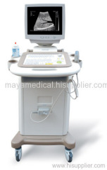
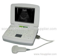
MA-2018 Standard ultrasound scanner (convex probe)
The system uses micro computer technology, Digital Scanning Converter (DSC), variable aperture, multi-section dynamic focusing, high dynamic, low noise, wide band pre-amplifier, log compression, TGC control, dynamic filtering, edge enhancement and frame correlation. Those embedded components allow a clear, stable and high resolution image.
.Display mode: B\B+B\B+M\M
.Gray scale: 256
.Image display: real time, frozen, amplified, B/W conversion, up/down conversion;
.Body marks: 10 types;
.Real-time, date, electronic focusing, free combination of focuses;
.Optional built-in pseudo-color processing mode; standard PAL-D and VGA video output; easily accessed to the equipments, such as the large screen monitor, videocassette recorder, video image printer etc.
.Soft keyboards and track ball to make operation faster, convenient and flexible;
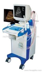
.Trolley structured; switch power is used without working frequency transformer; FPGA and SMT in the device make it high integrated, small volume and light weight.
.The device passed clinical verifications of safety and diagnosis validity in its manufacturing country.
Specifications
Standard probe: 3.5MHZ Convex;
.Detecting depth:≥ 220mm;
.Blind zone: ≤5mm;
.Display mode: B; B+B; B+M; M;
.Gray Scale: 256;
.Image amplification: ×1.0; ×1.2; ×1.5; ×2.0;
.Local zoom: 2 times;
.Electronic focusing: 4 focuses combination randomly;
.Depth shift: B, B+B modes real time shift;
.Resolution: lateral≤ 2mm; axial≤1mm;
.Frame correlation: B, B+B modes 3 level;
.Image reverse: up/down; left/right; black/white;
.Measurement: distance; circumference; area; heart rate; gestational week; fetal weight etc;
.Annotation: time; date; ID; age; sex; detecting depth; probe type; focus; frame correlation etc;
.Chinese-English inter-conversion;
.Cine-loop:186;
.Image load/storage; 20;
.Obstetric table: BPD; CRL; GS; FL; AC; HC; FW; EDD;
.Body marks: ≥10 types with probe position;
.Optional probe: 7.5MHz (linear); 6.5MHz Convex Trans-vaginal;